Cancer Chromosome Abnormalities Visualized in Living Cells
Cancer Chromosome Abnormalities Visualized in Living Cells
Researchers have observed for the first time how broken ends of chromosomes incorrectly reattach to each other—an abnormality common in cancer cells.
Chromosomes are structures inside our cells that are made up of tightly coiled DNA. The DNA contains genes, which instruct the cell how to make specific proteins, and non-coding sequences that orchestrate how those genes are used.

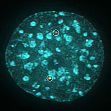
A chromosome translocation is visualized by breaks marked with differently colored fluorescent proteins (green, red). DNA is stained blue.Courtesy of NCI.
Humans normally have 23 pairs of chromosomes that package about 6 feet of DNA inside the nucleus of each cell. Many diseases and conditions are caused by abnormalities in the number or structure of the chromosomes. For example, having 3 copies of chromosome 21 rather than 2 copies causes Down syndrome.
Transfer of part of one chromosome to another, known as a chromosome translocation, can lead to cancer. Chromosome translocations are rare and are difficult to study. A team led by Drs. Vassilis Roukos and Tom Misteli of NIH’s National Cancer Institute (NCI) set out to understand how chromosome translocations form in living cells. Their findings were published in the August 9, 2013, issue of Science.
The researchers labeled different chromosomes in mouse cells with red or green fluorescent tags and then induced breaks in the DNA of the chromosomes. Using sophisticated imaging technology, they tracked whether the fluorescently labeled cut ends of the chromosomes reattached correctly to each other or in a mismatched fashion. For some experiments, they used time-lapse microscopy to simultaneously track several thousand cells for up to 24 hours.
The scientists found that most chromosome breaks reattached correctly, as cells have built-in repair machinery to fix DNA breaks. However, they were able to capture time-lapse images of rare translocations.
In further experiments, the researchers found that they could increase the likelihood of chromosome translocations by inhibiting key parts of the DNA repair machinery. In particular, when they blocked a protein called DNA-dependent protein kinase (DNAPK), a component of the machinery, translocations increased by almost 10-fold.
“Our ability to see this fundamental process in cancer formation was possible only because of access to revolutionary imaging technology here at the NCI,” Misteli says. “We can now finally begin to really probe how these fundamental features of cancer cells form.”
###
* The above story is reprinted from materials provided by National Institutes of Health (NIH)
** The National Institutes of Health (NIH) , a part of the U.S. Department of Health and Human Services, is the nation’s medical research agency—making important discoveries that improve health and save lives. The National Institutes of Health is made up of 27 different components called Institutes and Centers. Each has its own specific research agenda. All but three of these components receive their funding directly from Congress, and administrate their own budgets.


