New imaging technology may aid in early detection of breast cancer
New imaging technology may aid in early detection of breast cancer

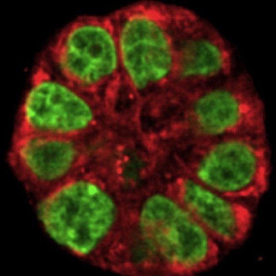
This kaleidoscopic image shows a live human mammary gland structure created by Purdue researchers working to develop a new imaging technology to determine a woman’s risk of developing breast cancer and advance research on methods for preventing the disease. Futurity reports:
The new imaging technique, called vibrational spectral microscopy, can be used to identify and track certain molecules by measuring their vibration with a laser. Whereas other imaging tools may take days to get results, the new method works at high speed, enabling researchers to measure changes in real time in live tissue, says Ji-Xin Cheng,[PhD] associate professor of biomedical engineering and chemistry.
By monitoring the same 3-D culture before and after exposure to certain risk factors, the new method enables researchers to detect subtle changes in single live cells.
Researchers say the work highlights the significance of engineering for the development of primary prevention research in breast cancer. Their findings are detailed in a paper(subscription required) published yesterday in Biophysical Journal.
By Lia Steakley
Stanford University Medical Center
Photo by Shuhua Yue at Purdue University
###
* Stanford University Medical Center integrates research, medical education and patient care at its three institutions – Stanford University School of Medicine, Stanford Hospital & Clinics and Lucile Packard Children’s Hospital.
** The above story is adapted from materials provided by Stanford University School of Medicine
________________________________________________________________


