Technique Forms Working Inner Ear Cells
Technique Forms Working Inner Ear Cells
Using an innovative 3-D culture system, researchers were able to coax mouse embryonic stem cells to form cells and structures seen in the inner ear. The technique could lead to deeper insights into inner ear development and disorders.
Specialized epithelial cells in the inner ear detect head movements, gravity and sound. Researchers know the general scheme of inner ear development but still have much to learn about how this specialized tissue forms. Deeper knowledge will be critical for developing novel therapies for hearing loss and balance disorders.

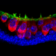
Stem-cell-derived sensory hair cells (in red) with hair bundles (green). Cell nuclei appear blue./ Courtesy of Indiana University.
Self-organized, complex tissues don’t arise naturally in traditional, flat culturing systems. To create a more lifelike environment, some scientists have recently created 3-D tissue culturing systems. By applying carefully timed developmental signals, researchers grew nerve tissues from embryonic stem cells floating in culture media. The cells aggregate and follow developmental pathways similar to those in the body.
A research team led by Karl R. Koehler and Dr. Eri Hashino at the Indiana University School of Medicine reasoned that a 3-D tissue culturing system might help to guide mouse embryonic stem cells to develop into the complex tissues and structures of the inner ear. The study was supported by NIH’s National Institute on Deafness and Other Communication Disorders (NIDCD) and other NIH components. It appeared online in Natureon July 10, 2013.
By experimenting with different signaling molecules and timing, the researchers were able to induce mouse embryonic stem cells to mimic the steps of inner ear development. The cells aggregated, and cell patches emerged within a week that bore a striking resemblance to the developing otic placode—the part of the embryo from which the ear develops. Tissue changes over the next few days continued to resemble inner ear development.
By day 16, the researchers saw cells resembling hair cells. These cells both underlie our sense of balance and help transform sound into electrical signals that the brain can understand. The hair cells generated electrical signals like those in the body. They also appeared to form special connections called ribbon synapses with adjacent neurons. These enable hair cells to pass their signals along to neural pathways leading to the brain.
“We were surprised to see that once stem cells are guided to become inner-ear precursors and placed in 3-D culture, these cells behave as if they knew not only how to become different cell types in the inner ear, but also how to self-organize into a pattern remarkably similar to the native inner ear,” Hashino says.
“The 3-D culture allows the cells to self-organize into complex tissues using mechanical cues that are found during embryonic development,” Koehler explains.
The method can now be used to explore the mechanisms involved in inner ear development. It can also provide a way to generate hair cells for drug discovery and other therapeutic testing.
By Harrison Wein, Ph.D.
###
* The above story is reprinted from materials provided by National Institutes of Health (NIH)
** The National Institutes of Health (NIH) , a part of the U.S. Department of Health and Human Services, is the nation’s medical research agency—making important discoveries that improve health and save lives. The National Institutes of Health is made up of 27 different components called Institutes and Centers. Each has its own specific research agenda. All but three of these components receive their funding directly from Congress, and administrate their own budgets.


