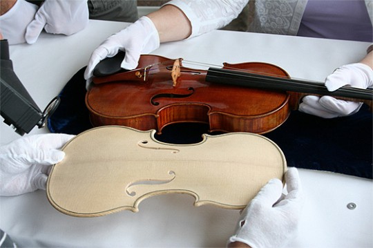
Stradivarius violin replicated through the magic of radiology
Stradivarius violin replicated through the magic of radiology
Radiologist (and amateur violinist) Steven Sirr, MD, and collaborators have created a reproduction of a 1704 Stradivarius violin using computed tomography (CT) images. According to a Radiological Society of North America release, here is how the team did it:
The original violin was scanned with a 64-detector CT, and more than 1,000 CT images were converted into stereolithographic files, which can be read by a computer-controlled router called a CNC machine. The CNC machine, custom-made for the project by Rossow, then carved the back and front plates and scroll of the violin from various woods. Finally, Waddle and Rossow finished, assembled and varnished the replica by hand.
“We believe this process of recreating old and valuable stringed instruments may have a profound influence upon modern string musicians,” Dr. Sirr said.
The original Stradivarius used is held by the U.S. Library of Congress. The team has posted a video of some of the CT imaging (.mp4) and of the CNC machine at work (also .mp4). A very good summary of the work is also available from the BBC.
By John Stafford
Stanford University Medical Center
Photo courtesy RSNA
###
* Stanford University Medical Center integrates research, medical education and patient care at its three institutions – Stanford University School of Medicine, Stanford Hospital & Clinics and Lucile Packard Children’s Hospital.
** The above story is adapted from materials provided by Stanford University School of Medicine
________________________________________________________________


