A Step Forward In Effort to Regenerate Damaged Nerves
A Step Forward In Effort to Regenerate Damaged Nerves
The carnage evident in disasters like car wrecks or wartime battles is oftentimes mirrored within the bodies of the people involved. A severe wound can leave blood vessels and nerves severed, bones broken, and cellular wreckage strewn throughout the body – a debris field within the body itself.

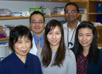
l-r: Weimin Liu, Jason Huang, Yi Ren, Samantha Dayawansa, Xiaowei Wang
It’s scenes like this that neurosurgeon Jason Huang, M.D., confronts every day. Severe damage to nerves is one of the most challenging wounds to treat for Huang and colleagues. It’s a type of wound suffered by people who are the victims of gunshots or stabbings, by those who have been involved in car accidents – or by soldiers injured on the battlefield, like those whom Huang treated in Iraq.
Now, back in his university laboratory, Huang and his team have taken a step forward toward the goal of repairing nerves in such patients more effectively. In a paper published in the journalPLoS One, Huang and colleagues at the University of Rochester Medical Center report that a surprising set of cells may hold potential for nerve transplants.

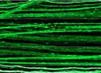
Nerve fibers created from DRG neurons
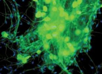
In a study in rats, Huang’s group found that dorsal root ganglion neurons, or DRG cells, help create thick, healthy nerves, without provoking unwanted attention from the immune system.
The finding is one step toward better treatment for the more than 350,000 patients each year in the United States who have serious injuries to their peripheral nerves. Huang’s laboratory is one of a handful developing new technologies to treat such wounds.

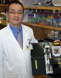
Huang with a device used to coax nerve cells to grow by stretching them
“These are very serious injuries, and patients really suffer, many for a very long time,” said Huang, associate professor of Neurosurgery and chief of Neurosurgery at Highland Hospital, an affiliate of the University of Rochester Medical Center. “There are a variety of options, but none of them is ideal.
“Our long-term goal is to grow living nerves in the laboratory, then transplant them into patients and cut down the amount of time it takes for those nerves to work,” added Huang, whose project was funded by the National Institute of Neurological Disorders and Stroke and by the University of Rochester Medical Center.

Thriving DRG cells
For a damaged nerve to repair itself, the two disconnected but healthy portions of the nerve must somehow find each other through a maze of tissue and connect together. This happens naturally for a very small wound – much like our skin grows back over a small cut – but for some nerve injuries, the gap is simply too large, and the nerve won’t grow back without intervention.
For surgeons like Huang, the preferred option is to transplant nerve tissue from elsewhere in the patient’s own body – for instance, a section of a nerve in the leg – into the wounded area. The transplanted nerve serves as scaffolding, a guide of sorts for a new nerve to grow and bridge the gap. Since the tissue comes from the patient, the body accepts the new nerve and doesn’t attack it.
But for many patients, this treatment isn’t an option. They might have severe wounds to other parts of the body, so that extra nerve tissue isn’t available. Alternatives can include a nerve transplant from a cadaver or an animal, but those bring other challenges, such as the lifelong need for powerful immunosuppressant drugs, and are rarely used.
One technology used by Huang and other neurosurgeons is the NeuraGen Nerve Guide, a hollow, absorbable collagen tube through which nerve fibers can grow and find each other. The technology is often used to repair nerve damage over short distances less than half an inch long.
In the PLoS One study, Huang’s team compared several methods to try to bridge a nerve gap of about half an inch in rats. The team transplanted nerve cells from a different type of rat into the wound site and compared results when the NeuraGen technology was was used alone or when it was paired with DRG cells or with other cells known as Schwann cells.
After four months, the team found that the tubes equipped with either DRG or Schwann cells helped bring about healthier nerves. In addition, the DRG cells provoked less unwanted attention from the immune system than the Schwann cells, which attracted twice as many macrophages and more of the immune compound interferon gamma.
While both Schwann and DRG cells are known players in nerve regeneration, Schwann cells have been considered more often as potential partners in the nerve transplantation process, even though they pose considerable challenges because of the immune system’s response to them.
“The conventional wisdom has been that Schwann cells play a critical role in the regenerative process,” said Huang, who is a scientist in the Center for Neural Development and Disease. “While we know this is true, we have shown that DRG cells can play an important role also. We think DRG cells could be a rich resource for nerve regeneration.”
In a related line of research, Huang along with colleagues in the laboratory of Douglas H. Smith, M.D. , at the University of Pennsylvania are creating DRG cells in the laboratory by stretching them, which coaxes them to grow about one inch every three weeks. The idea is to grow nerves several inches long in the laboratory, then transplant them into the patient, instead of waiting months after surgery for the nerve endings to travel that distance within the patient to ultimately hook up.
The first author of the PLoS One paper is research associate Weimin Liu, Ph.D. Other authors, in addition to Huang and Smith, are graduate students Yi Ren and Xiaowei Wang; post-doctoral associate Samantha Dayawansa, Ph.D.; undergraduate Adam Bossert; neurologist Handy Gelbard, M.D., Ph.D.; Jing Tong, M.D., formerly of the Huang laboratory and now a neurosurgeon at Hebei Medical University in China, and Xiaoshen He, M.D., a neurosurgeon at Fourth Military Medical University in China.
###
* The above story is adapted from materials provided by University of Rochester Medical Center
![]() ______________________________________________________________________
______________________________________________________________________


