Scientists trigger muscle stem cells to divide.
Scientists trigger muscle stem cells to divide
A tiny piece of RNA plays a key role in determining when muscle stem cells from mice activate and start to divide, according to researchers at the Stanford University School of Medicine. The finding may help scientists learn how to prepare human muscle stem cells for use in therapies for conditions such as muscular dystrophy and aging by controlling their activation state.

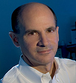
Thomas Rando
It’s the first time that a small regulatory RNA, called a microRNA, has been implicated in the maintenance of the adult stem cell resting, or quiescent, state.
“Although on the surface the quiescent state seems to be relatively static, it’s quite actively maintained,” said Thomas Rando, MD, PhD, professor of neurology and neurological sciences. “We’ve found that changing the levels of just one specific microRNA in resting muscle stem cells, however, causes them to spring into action.”
Rando, who is also the director of Stanford’s Glenn Laboratories for the Biology of Aging and the deputy director of the Stanford Center on Longevity, is the senior author of the research, published Feb. 23 in Nature. Postdoctoral scholar Tom Cheung, PhD, is the first author of the study.
Unlike stem cells in the blood or skin, muscle stem cells spend most of their lives nestled in the surrounding tissue. “They don’t do much most of the time,” said Rando. “They remain in a quiescent state for most of a person’s life. When you injure your muscle, however, they begin dividing to repair the damage.” Like all adult stem cells, each muscle stem cell becomes two daughter cells: one with stem cell properties, and the other that continues dividing to become mature muscle cells and fibers to replenish those that are damaged. Without such “asymmetric” division, the stem cells would quickly be depleted after injury.
Pinpointing exactly what calls the stem cells to begin dividing is an important first step to using them in human therapies. It’s also a key to understanding how muscles age and why they become less able over time to repair normal wear and tear.
“If you’re going to use muscle stem cells as a therapy for disease or aging, you want to be able to transplant cells that have the greatest potential to make new muscle in the recipient,” said Rando. “The quiescent state most closely resembles how they are in the body. If you allow them to divide in the lab before transplantation, they are not as effective. This microRNA may allow us to toggle the cells back and forth between the actively dividing and quiescent states.”
In recent years, microRNAs, which are only about 20 nucleotides long, have emerged from relative obscurity to claim a role as key regulatory molecules in the cell. They work by binding to the messenger RNA molecules entrusted to convey the information from DNA out of the nucleus to the protein-making machinery in the cell’s cytoplasm. Once bound, the microRNAs either target the messenger RNA for destruction or interfere with its ability to be translated into protein. MicroRNAs have recently been shown to be involved in controlling the fate of embryonic stem cells, so the Stanford team wondered whether they also played a role in adult muscle stem cells.
The team found that when they temporarily inhibited the function of all microRNAs in resting muscle stem cells from mice, the cells spontaneously activated and begin dividing. They also saw that fewer adult muscle stem cells remained in the tissue, and that mouse muscle fibers in which microRNA function was inhibited were less able than normal muscle to repair muscle damage over time.
Comparing the levels of expression of microRNAs between resting and activated muscle stem cells, the researchers identified 22 that were highly expressed in quiescent cells and markedly down-regulated in active cells. They homed in on one called microRNA-489 because its sequence is conserved across many species, suggesting it may be particularly important.
Muscles artificially induced to express much higher levels of microRNA-489 than normal were significantly compromised in their ability to repair damage — indicating that the muscle stem cells were unable to begin dividing. Conversely, those in which microRNA-489 activity was blocked had many actively dividing stem cells even in the absence of any injury.
“We were surprised that varying the expression levels of one microRNA could have such a profound effect,” said Rando.
When the researchers looked for the likely molecular target of microRNA-489, they found a protein called DEK that is known to be involved in cell proliferation and tumor growth. DEK expression levels are inversely correlated with those of the microRNA: when the microRNA level is high (in resting stem cells), DEK expression is low, and when the microRNA expression is shut off (in actively dividing cells), DEK expression is high — but only in the daughter cell that will continue to proliferate and become mature muscle fibers.
“The increase in DEK levels corresponds to many physiological changes,” said Rando, “and it’s asymmetric. Its presence confers the ability to divide on one daughter cell, while the other will remain a quiescent stem cell.”
In the future, the researchers will continue to look at the unique features of quiescent muscle stem cells, including those involved in normal aging.
“We’d like to understand the aging process at a very fundamental level,” said Rando. “That will allow us to move toward more therapeutic applications. Can we use what we’ve learned to convert old stem cells, which seem to have lost their responsiveness to activation cues, into young stem cells? Maybe the ability of old stem cells to exit the quiescent state is defective. We may one day be able to develop approaches that enhance tissue repair by enhancing stem cell function.”
In addition to Rando and Cheung, other Stanford researchers involved in the study include postdoctoral scholars Navaline Quach, PhD, and Ling Liu, PhD; graduate student Gregory Charville; undergraduate students Lidia Park and Bryan Yoo; and research assistants Abdolhossein Edalati and Phuong Hoang.
The research was supported by the National Institutes of Health, the Glenn Foundation for Medical Research and the Department of Veterans Affairs. Information about the Department of Neurology and Neurological Sciences, which also supported the work, is available at http://neurology.stanford.edu
By Krista Conger
Stanford University Medical Center
###
* Stanford University Medical Center integrates research, medical education and patient care at its three institutions – Stanford University School of Medicine, Stanford Hospital & Clinics and Lucile Packard Children’s Hospital.
** The above story is adapted from materials provided by Stanford University School of Medicine
________________________________________________________________


