Unlikely pairing of cardiologist, neurosurgeon gives young mother a 2nd chance
Brain Tumor Treatment Successful in Healing Rare Heart Condition
Unlikely pairing of cardiologist, neurosurgeon gives young mother a 2nd chance

Serendipity may have saved 32-year-old Jamie Arliss’ life. A rare tumor discovered in her heart had stumped cardiologists at the University of Rochester Medical Center. They wanted to remove the mass, but traditional techniques failed because of its size and location. A heart transplant was under consideration, that is, until a neurosurgeon suggested treating it like it was a brain tumor.
“This was an extraordinary tumor in the heart. So unusual, so rare that we couldn’t find any information about how to treat it anywhere in our extensive research,” said Christopher Cove, M.D., assistant director of the URMC Cardiac Catheterization Laboratories. The only information available came from autopsy reports when similar growths had been discovered, but no clues for how to treat it.
Fortuitous discussions between the cardiologist and a neurosurgeon introduced the idea of treating the tumor in her heart as they treat similar masses in the brain. The unlikely pair of specialists used liquid embolization – injecting a glue-like substance directly into the tumor — to stop the tumor in its tracks
“This was all new ground for us and we’re pleased that we’ve been able to stop the growth of that tumor without any negative impact on her heart,” Cove said. “We expect Jamie will be able to enjoy a good quality of life as she continues her recovery.”
Experts believe the Dec. 9 procedure was the first time this substance and technique was used in the heart.
> A Medical Mystery
Cardiothoracic surgeon H. Todd Massey, M.D., studied the images captured after it was discovered in fall 2008.
“It was nothing I’d ever seen before,” said Massey, senior transplant surgeon for the Program in Heart Failure and Transplantation. Initially, he was concerned that it was a cancerous tumor. However, a March 2009 biopsy debunked that theory, and Massey identified it as an arterio-venous malformation (AVM).
An AVM is a circulatory malfunction that can form anywhere in the body, though they occur often in the brain and lungs. When an AVM extends outside an artery or organ, creating a mass, it’s called a tumor. In Arliss’ case, it grew into a benign tumor the size of a golf ball along the lateral wall of Arliss’ heart.
Typically doctors try to remove the mass or cut off its blood supply through coil embolization or radiation therapy. Massey couldn’t remove it surgically because it would damage too much heart muscle. He suggested interventional cardiologists use a common coil embolization procedure to starve the tumor.
“That was unsuccessful because if you cut off one blood supply, it was able to find another one to let the tumor continue to grow,” Cove said, adding that the tumor was complex and deeply rooted in the heart.
And the life-threatening tumor kept growing, eventually reaching 3.5 centimeters and beginning to spread outside of the left ventricle and into the pericardium. The tumor was immense, since the average heart is about the size of a fist.
Doctors recommended Arliss consider joining the list for a heart transplant, a daunting prospect for the woman who wanted to continue her nursing education and raise a teen daughter with her husband, Scott.
> An Unusual Combination
Once cardiologists identified the mass as an AVM, they were stymied by how to treat it. There was nothing in the medical literature about AVMs in the heart. They reached out to colleagues across the country for advice and consideration. They shared the initial images of the mass, which were later published in the Journal of the American College of Cardiology in April 2010. While there was curiosity about the discovery, none of their peers offered suggestions for a curative therapy.

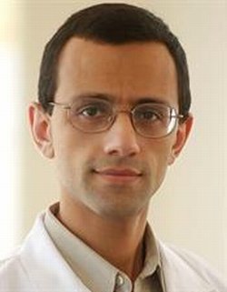
Babak Jahromi, M.D., Ph.D.
At the same time, neurosurgeon Babak Jahromi, M.D., Ph.D., temporarily moved his clinical suite into the cardiac catheterization center, during a renovation project. Cove and other interventional cardiologists observed Jahromi’s techniques for treating brain disorders, including AVMs.
Jahromi regularly treats cerebral AVMs by using a tiny catheter to inject Onyx, a glue-like substance, into the tumor to shut down the vascular growth and destroy the vessels.
Intrigued, Cove and Jahromi began studying the possibility of using the same substance to destroy the mass in Arliss’ heart.
“We decided to talk with the neurosurgeon next door because they have experience in treating these types of tumors in the brain,” said Cove. “We all use the minimally-invasive catheterization techniques to treat diseases and it was a great opportunity for cross fertilization between the two specialties.”
Jahromi knew there would be a learning curve working in a cardiac surgery instead of the Neurosurgery lab with a patient who is lying still under anesthesia.
“This was a completely different ball game for me,” said Jahromi, assistant professor of Neurosurgery, Imaging Sciences and Neurology. He typically uses the liquid embolization procedure to treat masses that are measured in millimeters, rather than centimeters or inches. The massive heart tumor required the use of 10 times the amount of Onyx – the thick, glue-like substance — than would normally be used in the brain. Onyx is coated with metal flakes that allow doctors to follow the flow of the liquid and see that it reached the target location. It hardens quickly an immediately shuts off the blood supply, destroying the mass.
“It was a beating heart that continues to move as you insert the catheter and deliver the therapy. And we did it all while the patient was awake. But that’s what the cardiologists do, so both of us working together we were a good combination,” Jahromi said.
Repeated imaging tests following the December 2010 procedure showed the Onyx filling the space where the tumor was, and no additional activity at the site. Now, it is an inert mass and it will no longer draw blood from Arliss’ heart. It could remain there for the rest of her life, though it will not impair her heart function, Cove said.
Doctors continue to monitor Arliss’ recovery and heart function, and will study what happens to the now-dead mass in her heart.
> Stumbling Onto a Diagnosis
Discovery of the tumor in Arliss’ heart came by accident two years ago. Arliss, a licensed practical nurse, was working at a medical practice near her hometown of Clyde, N.Y. At the time, the staff was learning to perform electrocardiograms (EKGs) and practicing on each other. When it was her turn, the test results were “a little odd and everyone said it was probably nothing,” she recalled.
Arliss had suffered progressive fatigue, as the tumor grew larger, reducing the capacity of her heart. She couldn’t exercise or complete tasks that required significant physical activity. She suspected something was wrong, but her doctors couldn’t find anything unusual.
“I always felt tired and short of breath, and I’d get chest pains sometimes. My doctor would order blood tests and they’d come back normal every time,” said Arliss, who is raising a 15-year-old daughter. “I quit smoking, started taking vitamins more regularly and paying attention to myself but it continued.”
After the unusual EKG, Arliss couldn’t ignore the results and she was persistent in finding out why. “I’m a worrywart and couldn’t let this go.”
Thus began a two-year journey that took many twists and turns as she found the road to recovery. Arliss is regaining strength and endurance and has returned to work in the medical office.
“We’re very pleased that Jamie will have no long-term effects from this procedure,” Cove said. “She’ll move on and enjoy a good quality of life from here.”
* The above story is reprinted from materials provided by University of Rochester Medical Center


