Brain Circuit Affects Visual Development
Brain Circuit Affects Visual Development
A mouse study revealed a circuit within the developing visual system that helps dictate how the eyes connect to the brain. The findings have implications for treating amblyopia, or lazy eye, the most common cause of visual impairment in childhood.

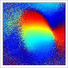
A retinotopic map shows the connections of the eyes to the binocular region of the visual cortex in a mouse brain. Amblyopia occurs when one eye is impaired and the other takes over this zone. /Image courtesy of Dr. Joshua Trachtenberg, UCLA.
Neural circuits can be shaped by experience—a quality called plasticity. One example of plasticity occurs when one eye becomes dominant over the other, a condition called ocular dominance. Under normal circumstances, both eyes send signals to a brain region called the binocular zone, giving rise to depth perception. As the brain develops, the eyes compete to connect within this zone. If one eye is hindered—for example, if it’s clouded by a cataract or if the eyes are positioned at different angles—it may lose space in the binocular zone to the other eye. Over time, the brain can develop a preference for the more functional eye at the expense of the other. Amblyopia is a condition in which the brain heavily favors one eye over the other, resulting in poor depth perception.
Competition in the binocular zone takes place during a limited time called the critical period. Patching the strong eye during this period can help correct amblyopia. But once the critical period closes—around age 7 in kids—the connections are difficult to change.
A research team led by Drs. Joshua Trachtenberg at the University of California, Los Angeles, and Xiangmin Xu at the University of California, Irvine, investigated plasticity and ocular dominance in the brain at a cellular level. Their work was funded by NIH’s National Eye Institute (NEI) and the National Institute of Neurological Disorders and Stroke (NINDS). The study appeared online on August 25, 2013, in Nature.
To induce changes in ocular dominance, the scientists temporarily patched one eye in young mice. After 24 hours, they removed the patch and recorded how the firing rate of binocular zone cells changed in response to vision through each eye.
The cells’ firing rates immediately dropped by half when vision was restricted to one eye, as expected. But over the next 24 hours, the cells responding to either eye—even the eye that had been temporarily patched—increased their firing rate back to the normal range.
The team took a closer look at this increased firing rate. They tested the possibility that binocular zone cells were getting more stimulation from other parts of the brain, but that wasn’t the case. Instead, the key turned out to be a brain circuit that normally inhibits the cells. When vision through one eye was impaired, there was less inhibition from cells called parvalbumin-positive basket inhibitory neurons, or PV cells. The weakened inhibition restored the binocular zone cells’ firing rate. Further experiments showed that this restoration of normal firing is the critical first step in the induction of ocular dominance plasticity.
The scientists also found that they could manipulate PV cells to re-start plasticity in older mice that were already beyond the critical period. The researchers note that if this circuit could be controlled in the human brain—for example, with a drug or implants—it could open the door to correcting amblyopia beyond childhood.
“Our study identifies a mechanism for visual development in the young brain and shows that it’s possible to turn on the same mechanism in the adult brain, thus offering hope for treating older children and adults with amblyopia,” Trachtenberg says.
###
* The above story is reprinted from materials provided by National Institutes of Health (NIH)
** The National Institutes of Health (NIH) , a part of the U.S. Department of Health and Human Services, is the nation’s medical research agency—making important discoveries that improve health and save lives. The National Institutes of Health is made up of 27 different components called Institutes and Centers. Each has its own specific research agenda. All but three of these components receive their funding directly from Congress, and administrate their own budgets.


